
EAP Publications | Virtual Library | Magazine Rack | Search | What's new
Join the Ecological Solutions RoundtableEAP Publication - 69
Microorganisms causing mastitis
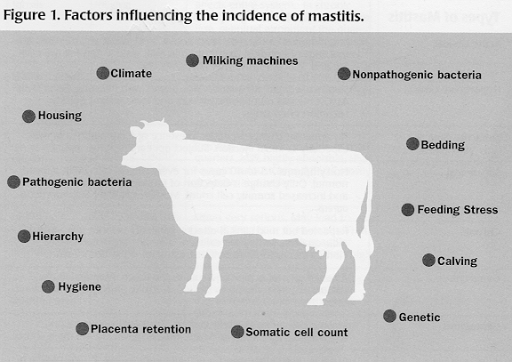
Factors contributing to mastitis
Physical and ethological factors
Counting and bacterial identification
Mastitis is a disease that affects a large number of dairy cattle throughout the world. A survey conducted in the major milk-producing countries indicates that each year clinical mastitis afflicts 15% to 20% of cows35. In Canada and the United States, it is thought that 50% of cows have one or more infected quarters. In Denmark, it is estimated that mastitis is the cause of 30% to 40% of veterinary interventions.
Using antibiotics is not an ideal solution. Other than the problems they cause with the milk (withdrawal for x days, contamination from antibiotic residues, problems associated with yogurt and cheese processing), antibiotics have not reduced the incidence of mastitis since their introduction (note A)37. Problems associated with resistance or even ineffectiveness are quite real in the case of mastitis caused by coliforms and Staphylococcus aureus15.
In organic agriculture, the use of antibiotics is neither normally authorized, nor desirable. There are however a large number of preventive and curative measures available to producers to deal with the problem. Mastitis control also entails a good understanding of the factors that encourage its incidence and the microorganisms that cause it.
Note A. The incidence of contagious mastitis has diminished through the use of antibiotics, but this has been paralleled by an increase in the level of environmental mastitis. Table 1 shows the difference between these two types of mastitis.
Since mastitis is a disease that has different levels of intensity and which may be caused by different organisms, there exists a complete jargon to describe the disease. Indeed, there are a number of types. It is therefore important to be able to recognize the different types of mastitis in order to decide what preventive measures to use as well as what treatment. To clarify the terms used throughout this article, Table 1 gives the definitions and characteristics of the different types of mastitis.
| Types of Mastitis | Characteristic Symptoms or Definition |
| Acute clinical | Inflammation of the teat, fever above 39 C, weak and dejected animal, lack of appetite. Drastic drop in milk yield. Often follows calving and, less seriously, after cow goes dry. |
| Hyperacute clinical | Swollen, red, painful quarter. Milk passes with difficulty. Fever over 41 C. Cow has no appetite, shivers and loses weight quickly. Lactation often stops. |
| Subacute clinical | No apparent change in udder, presence of flaky particles in milk, especially in initial ejection. Subject appears healthy. |
| Subclinical | No symptoms. 15 to 40 cases for every clinical case. Milk appears normal. Only change is detection of pathogenic agent in analysis and increased somatic cell count. Mostly caused by Staphylococcus aureus. |
| Chronic | Repeated but mild clinical attacks, generally without fever. Lumpy milk, quarters sometimes swollen. Quarter may become hard (fibrous indurations). Antibiotic treatments often do not work. |
| Gangrenous | Affected quarter is blue and cold to the touch. Progressive discolouration from the tip to the top. Necrotic parts drop off. Cow often dies. |
| Contagious | Mastitis caused by bacteria such as Staphylococcus aureus and Streptococcus agalactiae, of which infected cows are the main source. |
| Environmental | Mastitis caused by bacteria such as coliforms (e.g. E. coli), of which the main source is a contaminated environment, i.e. manure. |
There are a great number of microorganisms on and in cow udders. Watts52 identified 137 species and subspecies of microbes that can be associated with the mammary gland of the cow. Several of them are part of the normal flora and, with few exceptions, do not cause mastitis (note B). On the contrary, they may protect udders from infection caused by pathogenic bacteria.
Several other microorganisms may, however, cause infection in the mammary glands. The most common, those that cause about 90% of mastitis infections, are given in Table 2. There are contagious microorganisms and environmental microorganisms. Infected cows are the main source of contagious microorganisms, which survive and proliferate on the skin and on teat wounds. They consist of Streptococcus agalactiae, Staphylococcus aureus and Streptococcus dysgalactiae. Environmental microorganisms (Escherischia coli and other coliforms, Streptococcus uberis) do not remain on the teat. Rather, their presence indicates a high degree of contamination of soil, bedding, and water caused, particularly, by manure.
| Species | Main Source | Living Conditions | Propagation Factors | Symptoms | Preventive Treatment |
| Streptococcus agalactiae | Infected cows | Infected quarter and udder only | Using same rag for cleaning udders | Mild fever for about 24 hours | Wash udders after milking, reduces problem by 50% |
| Staphylococcus aureus | Infected cows | On abnormal udder and teat, milkers, vagina, tonsils | Transmitted by hands or rags, enters during milking | Often quite acute for a few days after calving. May be fatal. Quarter swells and turns purple. Quickly affects entire system. In chronic state, udder hardens, aqueous secretion, eventual atrophy of the quarter. Intermediate form with granular secretion. Milk hotter than normal. | Cull infected cows |
| Streptococcus dysgalactiae | Infected cows | Infected quarter, injuries | Pronounced swelling of one or more quarters. Milk highly abnormal. High fever in serious cases.Pronounced swelling of one or more quarters. Milk highly abnormal. High fever in serious cases. | ||
| Streptococcus uberis | Contaminated environment | On cow's skin, mouth, ground | Neglected udder washing, insufficient drying, lack of bedding, muddy yards | Pronounced swelling of one or more quarters. Milk highly abnormal. High fever in serious cases. Affects mostly dry cows and heifers. | Wash udders only, dry well with disposable paper towels for each cow. |
| Escherischia coli | Contaminated environment | Ground, bedding (sawdust and shavings), manure, water | Dirty calving stall, lack of bedding, inadequate udder washing | Often very serious. May lead to loss of quarter or even death. Thin yellow secretions, with granular texture resembling bran. Often high fever. | |
| Corynebacterium pyogenes | Certain insects | Humid valleys, wooded areas | Pronounced systematic reaction due to toxins caused by bacteria. Often more than one quarter affected. They become hard, produce thick smelly secretion like cheese and difficult to eliminate. Followed by abscess that bursts, releasing creamy pus, and tissue loss. |
To infect a quarter, a microorganism must first enter the quarter and the cow must be unable to get rid of it before it multiplies (note C). Following is a typical scenario that leads to mastitis infection.
1. Contact with the microbe: The number of microorganisms multiplies near the orifice (or sphincter) of one or several teats. This is where hygiene and milking habits play an important role in preventing microbes from entering the quarter.
2. Entry of microbe into the teats: Entry may be forced by the milking machine, particularly at the end of milking. Injured teats (injuries, keratin injured inside teat) or teats whose openings are too large may be easily invaded. This is where adjusting milking machines and preventing injuries is critical.
3. Immune response of the cow: the cow's first line of defense is to send white blood cells (leucocytes) to eliminate the microbes that have penetrated the teat. If the response is insufficient, the microbes multiply and the cow shows other immune responses such as fever. The effectiveness of the cow's immune system depends on many factors. This is also an area where farmers can do a great deal to ensure a good immune response.
Note B For example, Staphylococcus hyicus, Staphylococcus epidermis and Corynebacterium bovis.
Note C It is possible that some microorganisms enter the udder from other parts of the body, but this possibility is still under debate. It therefore must be assumed that the main entry of microbes is the teat.
Mastitis is a difficult problem to comprehend because it is a disease caused by many factors. Microorganisms are responsible for the infection, but for them to enter the mammary glands and establish themselves to the point that they cause an infection, a multitude of factors may be involved. There are many such factors (e.g. hygiene, housing, climate, milking machines, feed, genetics) acting simultaneously. It is even more difficult to generalize about the relative importance of each one, as certain factors affect certain microorganisms in particular.
Klastrup and his coworkers21 estimate that 25% of the susceptibility to infection is attributable to environmental factors, 20% to genetic factors, and 50% to herd management.
Climate
Climate may have a direct or indirect influence on the onset of mastitis. Older authors7,43 insist a great deal on the fact that exposure to intense cold, draughts, excessive humidity or heat predisposes cattle to mastitis.
Just as they foster our colds, rapid changes in temperature can encourage mastitis. Research on how temperature influences the incidence of mastitis indicates that temperature extremes interact with other factors to cause mastitis but rarely will temperature alone cause the disease21. Temperature extremes may also affect somatic cell counts. Therefore the incidence of mastitis increases with extreme temperatures. In Florida, a higher rate of clinical mastitis was observed three years in seven during very hot periods28.
A particular type of mastitis often called summer mastitis, is caused by biting insects that contaminate the udder with the bacteria Corynebacterium pyogenes and other anaerobic bacteria. The frequency of this type of mastitis varies according to the regions, with humid valleys being most susceptible.
Climate may also have an indirect influence. For example, muddy conditions outdoors caused by abundant rainfall will increase the number of microorganisms and thus increase risks of infection.
Housing
The mere fact of keeping cows indoors increases the incidence of mastitis. When cows are inside, the risk of udder injury increases. There are also microorganisms whose populations are less concentrated outdoors. In Australia, where cows go indoors only to be milked, mastitis caused by coliforms is rare.
Although the question is often debated, it would appear that mastitis is less common with loose housing systems than with tied housing systems. It might be thought that mastitis is more frequent with loose housing because the microbes are easily transmitted from one cow to another. However, cows are usually happier in loose housing. They are also less likely to injure themselves or come into contact with soiled bedding and so are less subject to mastitis. The social adjustment of cows within the herd is also clearer with loose housing. According to a Serbian study27, there are 27% less cases of subclinical mastitis and 42% less cases of clinical mastitis in loose-housed herds than in herds kept tied.
In tied housing, there is more likelihood of mastitis with some types of stalls. The longer and wider a stall, the more the cow can move, thereby reducing the number of injuries and the incidence of mastitis19. The greatest disadvantage is that the animal's vertical movements are restricted, particularly when getting up or lying down. Partitioning between the stalls reduces the incidence of mastitis as a result of abrupt movements of neighbouring animals and the likelihood of teat tramps.
Quality of indoor air
Draughts, excessive humidity and frequent changes in temperature in a barn are factors that lead to increased incidence of mastitis. As seen in previous pages, indirect effects on an animal's immunity have not been seriously studied. However, effects on the concentration of pathogens in stables has been more so. For example, the bacteria Klebsiella pneumoniae causes more infection when relative humidity is low48, while the number of infections caused by E. coli does not vary with humidity changes.
Bedding
Whether with loose or tied housing systems, bedding plays an important role in the incidence of mastitis. This is easy to understand when considering the mastitis-infected milk that reaches the ground, the humidity that favours the development of microbes on bedding and that cows often spend 14 hours out of 24 in contact with their bedding. In an experiment where cows were housed with and without bedding, the level of mastitis infections doubled where there was no bedding. Inadequate bedding in loose-housed herds, particularly large herds, may lead to serious situations in the case of contagious mastitis.
Different materials used as bedding may affect the growth of different microorganisms. Straw is generally the most recommended bedding. There is less rapid development of pathogenic microorganisms with chopped oats straw and cedar sawdust than with newsprint4. Chopped straw, however, is more favourable to Klebsiella than sawdust16. Sawdust and shavings, particularly if heated, encourage the rapid development of coliforms in general and are often responsible for "epidemics" of coliform mastitis36.
Stress
The more an animal is stressed in its environment, the less efficient its immune system is, and the less it can resist microbial infestations. Therefore, the more stress there is, the greater the chance of mastitis10. Giesecke10 has even demonstrated that stress affects the integrity of intramammary cells, which is yet another factor contributing to mastitis. The following are some sources of stress:
- Excessive density of animals. Proximity of cows encourage microbial exchanges and tense relations between animals;
- Irregular management, unpredictable behaviour on the part of the farmer;
- Noise;
- Stray voltage.
Recently there has been a lot of research done on how hereditary factors influence susceptibility to mastitis. The different dairy cattle breeds are not equally susceptible to mastitis. High yielding cows are more likely to be affected. Selective breeding that focuses solely on milk production is undoubtedly an important factor in higher rates of mastitis. According to different sources, hereditary factors account for 12% to 20% of susceptibility to mastitis in a single breed.
Cows selected for several traits have a higher somatic cell count (better immune response), requiring almost two times less treatment, and their milk is thrown away half as often as the milk of cows selected for one trait, although the latters produce more milk49.
Genetically, there is a correlation between the percentage of milk fat and the incidence of clinical mastitis. The more a line of cows gives fat milk, the more it will be susceptible to mastitis. It is therefore important to not select only on this basis.
Despite several serious studies on the subject, the links between diet and mastitis still raise questions in scientific circles. Two practices that increase the risks of mastitis are rapid changes in diet and excess or imbalance in the different components of rations.
Nitrogen and proteins
Excessive nitrogen or protein in feed is often mentioned as one of the factors causing mastitis. According to a Danish study25, there is no definitive link between protein content in diet and the incidence of mastitis. However, there is more evidence regarding the harmful effect of nitrogen that is not in a protein form (e.g. urea and ammonia) on the incidence of mastitis.
Nonprotein nitrogen (NPN) is particularly hard on white blood cells, or leucocytes, which protect the udder. Abrupt changes to rations based on NPN-rich high moisture corn or alfalfa silage should be avoided. Increases, however modest, of ammonia in the blood has repercussions on metabolism. If such rations are used, sufficient fibre should be included to feed microorganisms in the rumen that will convert the nonprotein nitrogen into bacterial protein.
According to an experiment realized in Germany8, there is a significant relationship between the level of urea in the blood and bacterial colonization in the udder. In another experiment, the addition of urea to rations increased susceptibility to infection and increased the number of infections by more than 16%45. The effect on the immune system is particularly evident when the urea is given in large quantities (over 18Og/day more than nitrogen requirements)2.
Concentrates and energy
It is recommended that reduced quantities of concentrates be given to a cow with mastitis. It appears this is also true for preventing mastitis, according to a German study22 conducted on 1038 first lactation cows and 572 cows of the following lactations. When the cow rations contained 25% concentrates rather than 40%, the incidence of mastitis was 7% compared to 36% for first lactation cows and 19% in comparison to 37% for other cows.
The same study also compared different energy levels in rations. A high energy content in rations increased the incidence of mastitis in first lactation cows whereas it had the opposite effect on the other cows.
Calcium-phosphorus ratio
An inadequate calcium to phosphorus ratio in rations results in problems with milk fever at calving40. In large herds, up to 50% of animals lacking calcium in their rations will develop coliform mastitis a few hours after calving. This hypocalcemia generally results from an inadequate calcium to phosphorous ratio in rations during the dry period. See page XX for more information on rations to give during the drying off period.
Silage and hay
Poor quality silage has a very negative effect on the immune system. The overheated proteins and sugars may kill the white blood cells protecting the udder. Cows fed with hay and grain have greater resistance in every way to several pathogens than cows fed with silage38. In some cases, Pseudomonas and Proteus are the only microorganisms that survive the high temperatures produced in silage. Although rare, silage contaminated this way may then be a source of mastitis caused by these types of organisms. Mouldy hay and mycotoxins also harm white blood cells and therefore weaken the immune system.
Alfalfa and other legumes
Legumes, particularly alfalfa, contain estrogenic substances whose concentration varies depending on plant maturity. Turning legumes into silage does not reduce their estrogenic properties. Through a physiological mechanism, that is still not well understood, these external estrogenic substances (that is, they are not produced by the cow herself) tend to foster mastitis. Several studies indicate that alfalfa added to the rations of cows with chronic mastitis exacerbates the infection. What is important is not to feed silage with a high legume content to heifers. This estrogenic intake encourages premature development of the udder and increases the incidence of environmental mastitis21.
Selenium and vitamin E
In the last ten years, several researchers have looked into the use of supplements and the role of selenium and vitamin E in the prevention and treatment of mastitis. Maintaining an adequate level of selenium in the organism helps prevent mastitis, reduce the severity of infection and causes it to last for a shorter period of time. Selenium serves to reinforce the immune system response by increasing the release of leucocytes and increasing the efficiency of phagocytes9. Selenium and vitamin E work together in the organism. Thus, a vitamin E supplement of 1000 lU/day alone reduces the somatic cell count but not the incidence of mastitis3.
With both selenium and vitamin E supplements, it can be expected that infections will be reduced by 42% at calving, by 59% for the entire duration of the infection and by 32% for clinical mastitis. The role of selenium is considered to be most significant in the case of subclinical mastitis30.
Selenium supplements may play a particularly significant role in cases of mastitis caused by E. coli. For example, cows that are given a selenium supplement of 0.35 mg/kg dry matter are better able to resist mastitis caused by E. coli24. The duration of this type of mastitis is even shorter when cows receive 2 mg of selenium per day per kilo of ration9.
Recommended blood levels are 0.2-1.0 g/ml for selenium and more than 4 g/ml for vitamin E. Rations should provide 3 mg of selenium per day in the case of dry cows and 6 mg per day for producing cows. Rations should provide 1000 IU of vitamin E per day for both categories of cow44. Supplementation with vitamin E has a greater effect on dry cows than on lactating cows, where a good part of vitamin E supplements is eliminated in the milk.
Important: It is not useful and even harmful to give only large doses of selenium (that is, without vitamin E), because the effect can be toxic. A selenium dose of 16 mg/day results in higher levels of mastitis unless vitamin E supplements are administered at the same time53.
Silica
Finnish researchers34 noted that the level of silica in mastitis-infected milk was only 0.39 mg/litre whereas it was 0.81 mg/litre in normal milk. Also, the level of silica in the blood serum of cows infected with mastitis is 1.02 mg/litre rather than 1.63 mg/litre for uninfected cows. Silica, whose role is similar to selenium, has a marked effect on the formation of free radicals, lipid peroxidation and macrophage activity. The silica content in rations may be increased by giving high silica-content feed like cereal straws.
Other nutritional factors
Vitamin A deficient rations reduce immunity. An Italian researcher experimented with vitamin A and beta-carotene supplements to control mastitis11.
According to Katholm18, iron also plays an important role in the prevention of mastitis. It is linked to the protein lactoferrin.
Needs of the calf
The famous animal phytotherapist Juliette De Baïracli-Levy6 believes that preventing cows from benefitting from the pleasure and stimulation of nursing their calves is one of the principal causes of mastitis. With suckling calves, Levy makes a distinction between the "psychological" and physical factors.
It is often observed that cows separated from their calves shortly after calving look and call for them. The question of course is whether cows experience a difficult "emotion". If this hypothesis is accepted, however, it would suggest that cows who find the separation more difficult, develop mastitis more easily.
On a physical level, the frequency of calf suckling is greater than cow milking. Microorganisms that invade a quarter have very little time to develop. Should cows therefore be milked more often at the beginning of lactation? Slavic researchers47 have observed that the duration and frequency of mastitis was lower during the first two months following calving with cows that nursed their calves for six to ten days as opposed to one hour, two days or four days.
Herd hierarchy
With loose housing or pasturing, a hierarchy is created within the herd, a phenomenon that is even more apparent in goats than in cows. It is possible that the least dominant members of the herd, who are often harassed by the others, have a greater tendency to develop diseases. The advantage of loose housing is that the hierarchical relationships between members is clear. Cows in tied housing can be quite stressed when they find themselves in an exercise yard where relationships between cows are not clear.
Uterus-mammary glands
It has been demonstrated that cows who retain their placentas have mastitis more often that those who do not14. The risk of developing mastitis is increased threefold42. Mastitis is clearly associated with placenta retention in the case of mastitis caused by Actinomyces pyogenes according to German researchers55. This type of mastitis represents 17% of cases in Germany. Mastitis that appears within the two months following calving is often linked to uteruses that are not cleaned properly. Discharges of purulent matter dirties the tail and rear end of the animal, and the ground, which favours environmental contamination and, subsequently, the udder. Some veterinarians venture further by saying that the reproductive organs may serve as reservoirs of infection. The pathogens thus travel through the blood to the mammary glands. At any rate, beware of placenta retentions!
Rumen-mammary ulands
The rumen is a very important organ of the cow, and the health of the other organs often depends on it. When acidosis occurs in the rumen (too much grain in rations for instance), it creates conditions that foster bacteria like Streptococcus bovis and eventually yeasts like Candida albicans. Therefore, although rare, the toxins from these substances can travel throughout the system and favor gram-positive bacteria that attack the udder54.
The approach of most laboratory studies is to consider factors in an isolated manner. In mastitis research, there are numerous studies based on farm practices. These studies rely on questionnaires addressed to farmers, analysis results, etc. One such study that was particularly original was conducted in eastern Ireland46, and integrated human factors with herd management factors. The results are given in the table below. It should be noted that the farmer's age and teat washing did not come out as determining factors in the incidence of mastitis during the study.
| Characteristic | Associated factors |
| Low somatic cell count | Geographical position of the farm, treatment of dry cows, replacement cows produced on the farm, positive attitude towards milking, family enterprise. |
| High somatic cell count | Small herd, irregular maintenance of milking equipment, lack of bedding on cement floor, udder washing of dirty cows only, little ambition. |
| Low bacterial count | Treatment of dry cows. |
| High bacterial count | Tied housing, obsolete milking equipment, short withdrawal period after antibiotic treatment, slight tendency to seek out information. |
| High milk yield | Average herd, treatment of dry cows, average tendency to seek out information, elimination of cows that are too susceptible. |
| Low milk yield | Lack of hot water at milking, use of one cloth only for all cows, infrequent meetings with other farmers, strong will to continue traditional family farming, no vacation. |
To diagnose mastitis, it is necessary to learn how to distinguish between the symptoms of the various types of mastitis infection (see Tables 1 and 2 above). The key points to remember are as follows:
Monitor the milk: routine examination of the milk using a filter cup to extract the first three squirts before washing (before milking) is undoubtedly the best way to diagnose mastitis. The presence of lumps, flakes, blood, etc. must be watched for. Milk that is hotter than normal may be a good indication of a Staphylococcus aureus infection.
Palpate the udder: particularly after milking, when it is easy to detect swelling, and fibrous, hard or injured tissue.
Be attentive: to other more evident signs such as fever, redness, etc.
Since these symptoms are often absent, particularly in cases of subclinical, subacute or chronic mastitis, only half of all mastitis infections, at best, can be detected through observation. Some tests may therefore also be useful, notably cell counts, bacterial identification and the California Mastitis Test (CMT).
All producers registered in a dairy record of performance are familiar with somatic cell counts. Sometimes wrongly referred to as white cell counts, this type of test includes all somatic cells, including white cells (or leucocytes) and epithelial cells. When swelling occurs, the cow's immune system reacts by sending leucocytes to destroy the foreign bodies. The somatic cell count in the milk may thus indicate if a cow is fighting infection.
Cell counting came into use largely to ensure that milk from a given herd was fit for human consumption. It is also a highly useful test for mastitis detection, albeit lacking in certain respects.
First, cell counts do not distinguish between leucocytic and epithelial cells. For example, for normal milk with a cell count of 50,000 cells per ml, there may by 20% leucocytes and 80% epithelial cells, whereas mastitis infected milk with a cell count of over 500,000 cells per ml, contains 90 to 95% leucocytic cells, although this ratio may also be quite different. It would be easier to determine if a cow was fighting infection if only the leucocytic cells could be counted. Since the count does not make the distinction, it is difficult to interpret the results of a count, especially with average numbers. With high cell counts, the so-called "millionaire" cows, the diagnosis is clearer and is indicative of mastitis.
Second, there is tremendous variation between the number of somatic cells with or without mastitis. The number of somatic cells in milk is generally higher in summer months, higher at the beginning and end of lactation and increases with the age of the cow. It also depends upon the genetic history of the cow and bull13. Moreover, an increase in somatic cells may be linked to a functional disorder of the reproductive organs. Even with a sampling and count that are properly carried out, a difference of 25% from one day to another can be expected without the circumstances of the herd having changed29.
Third, cows in one herd react differently to infection than cows from another herd. For example, Natzke29 reports that the average cell count of milk from quarters infected by Staphylococcus aureus was 6,700,000 in one herd and 900,000 in another. In the same study, the non-infected quarters had a cell count of 600,000 in one herd and 150,000 in the other.
Finally, when a cell count is high, the animal or the milk generally shows obvious symptoms of mastitis. The test therefore tells us nothing more than if we had been attentive.
Despite these shortcomings, the somatic cell count remains an important and practical tool for measuring the general health of a herd or individuals. Natzke29 believes that cell counting is particularly useful for evaluating the long-term health of a herd. Observing cell count trends from one year to the next serves to evaluate if any progress has been made, or if the situation is stable or deteriorating.
Counting and Bacterial Identification
In several states and provinces, bacterial counting in milk is done monthly for all dairy farms. It consists of evaluating the size of populations of different microorganisms in milk samples.
The identification of microorganisms present in the milk is sometimes useful on the farm to determine exactly what species of bacteria are responsible for the infection. This is the case for example when several cows have the same symptoms. These tests are done at government's pathology laboratories.
Clean milking habits are important to avoid the spreading of germs or their proliferation. The purpose of hygiene is to prevent the transmission of germs from one teat to another on one cow or from one cow to another.
There is no need to get overly excited about germs... but why provoke them?
Pasteur admitted at the end of his life that "the terrain is everything, the microbe is nothing", meaning that pathogenic microorganisms could not cause disease in a healthy animal or plant (well fed, etc.). Although optimum health is always the ultimate goal, it is not always easy in to attain in herd management. Therefore, in the meantime, a little hygiene can't hurt!
Udder washing
Washing the udder is hygienic and it has a stimulating effect on milk flow. Adequate washing is especially important to prevent environmental mastitis, caused by coliforms and other microbes from contaminated environments. Badly washed udders contribute to the transmission of microbes rather than to their destruction.
According to Pankey33, the lowest bacterial count in milk is obtained by washing the udder in the following fashion:
- Using individual moist paper towels, wet and wash the teats only. Wetting the udder and the teats results in more bacteria getting into the milk than if only the teats are wet.
- Dry with individual paper towels.
Note that teat dipping before milking in addition to drying off does not give better results than drying alone, and it increases the risks of contamination of the milk by disinfectants.
Udder washing This is a recipe for washing udders used by Daniel Lapointe, an organic dairyman from Quebec. Although the antiseptic value of this formula has never been scientifically tested, he has been using it for many years with good results. It consists of mixing 13 litres of hot water, 1 drop of pine oil, 1 capful of peroxide and 1 ounce of clay. |
Foremilking
Removing a little milk by hand before machine milking serves to stimulate milk letdown and to obtain a milk sample containing a high microbial count. A filter cup is used to detect abnormal looking milk (lumpy, etc.).
Milking order
It is important to milk infected cows last. If possible, milking order should be as follows: first lactation cows, normal cows, cows with a high cell count and then infected cows.
Other measures during milking
It is important to milk completely. With modern milkers, the risks of forcing the entry of microbes at the end of milking greatly diminishes, as long as they are well adjusted. The chances of bacteria entering the udder can be reduced by diminishing the amplitude of the vacuum changes and the vacuum change speed on the teats12. To do so, a good vacuum reserve and appropriate piping are necessary. Furthermore, the milker must not slip on the teats and the milkers must be removed carefully.
Risk of infection may be diminished if milking is finished by hand, although not realistic for an entire herd. De Baïracli-Levy6 even suggests massaging the udder after milking and hitting it up and down in the same way that calves do.
It is important to milk twice a day, even with cows that do not produce a lot. The longer the milk remains in the udder, the greater the risk of infection. The first squirts of milk must not go on the ground as this will contaminate the bedding and floor.
Postmilking teat dipping
Using a disinfectant teat dip after each milking is a means of diminishing by about 50% the risk of infection by contagious microorganisms like Streptococcus agalactiae and Staphylococcus aureus. Teat dipping prevents populations of these microbes from developing sufficiently between milkings. Teat dipping also discourages flies.
It is important that the teat dip contain up to 10% of emollients to increase the suppleness of the teats: oils, glycerine, lanoline. Healthy supple skin is an extra assurance against entry of bacteria to the udder. Staphylococcus aureus do not persist on healthy skin.
| Teat
Dip This is another recipe by organic dairyman Daniel Lapointe. You simply mix four litres of water, 5 ml of lavender oil, 5 ml pine oil, 2 ml eucalyptus oil, 12 ml cottonseed oil (available in drugstores) and 5 ml methylene blue. |
Cleaning equipment after milking
It is obviously important to clean and disinfect equipment after milking. Cider or corn vinegar and peroxide are used by some producers as alternatives to phosphoric acid and chlorine.
Milking Method Recommended in Quebec
Material:
Filter cup, hot water at 55 C with disinfectant, paper towels, pails for used towels and container for teat dip.
Milking Method:
1. Collect first three squirts of milk in a filter cup.
2. Soak towel in hot water with teat dip disinfectant, and wash each teat, using the other side of the towel to wash the teat sphincters.
3. Using another towel, wipe off the four teats and then with the other side of the towel massage the four teats by rubbing vigorously in a circular motion: 0-3 months of lactation = 10 times; 4 -7 months lactation = 15 times; 8 months lactation and more = 20 times.
4. Install milking unit on each cow, without losing vacuum.
5. Shut off the vacuum when milk flow is insufficient, remove the milker claw and immediately wash the teats.
Note: Only 15% to 20% of cows need to be milked dry. In this case, press gently on the milker claw while massaging from bottom to top the quarters that still contain milk.
Indoors
Abundant bedding prevents injury to the udder, limits exposure to cold damp floors and limits contact of the udder with manure. A minimum of 3 kg of straw per day per animal must be used (about one ton per cow per year). It is better to use a little bedding often, rather than a large quantity less often. Straw is preferred. Adding lime to the bedding can help in a stable where environmental mastitis is a problem but can also irritate the udder, the teats and the lungs when airborne.
It is important to prevent the cows from injuring their udders. The floors should not be slippery when the cows are let outdoors and there should be separators between the cows. Disinfecting the stable twice a year is a good practice.
Outdoors
There should not be any mudholes around the buildings or any place the cows have access to. Along the same lines, watering areas should not be allowed to become mucky. Ideally they should be located on higher ground or gravel or cement platforms used under the drinking areas.
There should be no barbed wire left lying around or in areas where the cows could injure themselves.
Overpopulation in the stable and fields, particularly with loose housing, should also be avoided, as it increases stress on the animals and risk of contagious mastitis being transmitted.
Changes in feed must be done slowly. Excesses must be avoided, particularly concentrates and nonprotein nitrogen feed (e.g. alfalfa and high moisture grain corn silage). A 1.4 to 1.8 calcium to phosphorous ratio must be maintained, even during the dry period. Selenium and vitamin E supplements may be a good choice if the ration does not provide the necessary minimum. For more details, consult the section on nutritional factors on pages x, x and x.
Replacement: Do not buy infected animals; have them tested before purchasing them and examine the udders. Research in several countries has demonstrated that up to 50% of purchased cows have subclinical infections37. It is better to buy only heifers (heifers generally do not have mastitis) or produce your own replacement animals. In any case, heifers should not be suckled as this breaks the teat seal and thus facilitates entry of microorganisms that can cause mastitis at calving.
Culling: Cull animals that are severely or repeatedly affected by mastitis. Cows with injured teats that do not heal should be put at the top of the list of animals to cull. They are up to 10 times more likely to contract mastitis. Cows that maintain a high cell count during all lactations should also be culled.
It is well known that mastitis often affects cows that have recently gone dry. These animals should not be overfed, particularly during dry periods. First lactation cows in particular must also be supervised since they are twice as likely to develop mastitis during the dry period than the others29.
In conventional agriculture, the merits of treating dry cows with antibiotics is often considered as one of the most effective methods along with the postmilking teat dip to reduce incidence of mastitis. The significance of this for organic agriculture is that the dry cow must not be overlooked.
Change in feed at drying-off is important. Following are the three steps:
Post-lactation (7 to 14 days): give a reduced diet of fibrous and poor hay to provoke rapid drop in milk flow and to stimulate the rumen. Drinking water must be drastically reduced. Some organic farmers give 4 drops of sage or menthol essential oil and charcoal two times a day to cut production at this stage.
Dry (30-90 days): diet is made up mostly of roughage with a good energy-protein and mineral balance.
Pre-lactation (7 to 14 days before calving): moderate quantities of energy-rich concentrates are added to a balanced roughage ration.
Use of the teat dip before and after the dry period (i.e. 15 days before calving and 15 days after the dry period) may be beneficial in herds where clinical mastitis is common32.
The following curative measures are particularly for clinical and chronic mastitis. There is a vast range of curative methods that may be used as an alternative to antibiotics: homeopathy, clay therapy, phytotherapy, etc. The advantage of homeopathy over antibiotics is that milking may be continued. The other alternative products used must not go in the bulk milk because tests for detecting antibiotics in milk may react positively to certain products like some essential oils.
When a treatment is being administered it is important to modify other practices:
- Infected cows must be fed prudently. Concentrates must be reduced and extra fibres and laxatives must be included. In cases of clinical mastitis, Eckles7 recommends reducing grain rations by one third as soon as symptoms appear and until they disappear;
- A purge must be given (except with homeopathic treatments);
- Avoid exposing infected animals to cold and draughts;
- Milk gently by hand 3 to 6 times a day.
Extreme caution must be used with products that are injected into the teat. It is quite difficult to make a «clean" injection in a contaminated environment such as a dairy barn without provoking a new contamination in the quarter.
In all cases, it is important to immediately contact a veterinarian if there is no rapid improvement.
Clay has several therapeutic properties. It has proved efficient as poultice to treat inflammation caused by mastitis on account of its high absorbency. To prepare a clay dressing, the clay is mixed with a liquid. Some producers use room-temperature water, while others use olive oil. A good compromise is to use half water, half oil, with the oil giving a more elastic consistency to the paste. The final result must be fairly liquid while adhering well to the udder.
When using water, it can be allowed to penetrate slowly into the clay without mixing. The container should be covered with a cloth and set out in the sun. The oil mixture on the other hand must be mixed. In both cases, a wooden spoon must be used and ideally the container should be a non-reactive material like porcelain or glass. The therapeutic effect of the paste may be increased by adding 2 to 3 drops of pine or thyme oil for every two litres of mixture.
The clay poultice must be spread over the infected parts of the udder after milking. The clay may be removed once dry and replaced 2 to 3 times a day by a new application. The dressing may be left on all night after evening milking.
In the case of the mixture containing oil, the mastitis has healed when the udder stays oily after the dry clay has been removed. This treatment should produce results in two to three hours in the case of acute mastitis, 4 to 6 hours with less serious forms and two to three days with chronic mastitis. It the treatment does not appear to be having any effect after this time, other measures must be considered.
The following information is for general consultation only and should not be considered as a replacement for professional advice from a homeopathic veterinarian.
Prevention
Preventive homeopathic treatments are administered using nosodes on an entire herd rather than on individual animals. Nosodes are fragments of pathogenic cells that increase the immune response. Nosode preparation is established according to the species of bacteria causing the problem in the herd. MacLeod23 recommends a nosode of 30 dilutions in liquid form administered by the mouth or in the herd's drinking water.
Complexes
An alternative to nosodes that is particularly effective for subclinical mastitis is to use in conjunction homeopathic remedies that have proven effective against mammary gland afflictions. MacLeod (1981) recommends the following combinations as multiple use remedies: Belladonna, Bryonia and Urtica Urens; Phytolacca and Sulphur, Silicea and Carbo vegetabilis.
Most homeopathic laboratories (see list of useful addresses on page XX) offer specific complexes for mastitis. The advantage of these complexes, which heal three out of four cases, is that they are easy to use. Some practitioners do not recommend the use of complexes on animals that are to be kept for several years because these substances tend to "confuse" the immune system.
Homeopathic treatments are administered through the mucous membrane. Homeopathy is less effective when the infection is caused by injury to the teat.
Specific treatments
More specific homeopathic remedies are selected according to the symptoms, such as lumpy milk, edema type, etc. Those used by two famous homeopathic veterinarians, the British MacLeod and the French Quiquandon, are given in Tables 4 and 5.
| Homeopathic Remedy | Symptoms | Dose |
| Belladonna 1 m | For acute postpartum mastitis. Udder very hot and red, painful to the touch. Animal is hot, and pulse is quick and strong. | 1 dose every hour. 4 doses. |
| Aconitum 6 x | Routine treatment for all acute cases, particularly those that develop rapidly after exposure to cold dry wind. Relieves tension and anxiety. | 1 dose every half-hour. 6 doses. |
| Apis Mellifica 6c | Indicated for first calving, heifers with edema of and around the udder. Mammary vein is swollen. | 1 does every 3 hours. 4 doses. |
| Bryonia Alba 30 c | Indicated for swollen and very hard udders. Pain is less intense when pressed. Animal is often Iying down. Especially good for chronic cases with fibrosis. | Acute cases: 1 dose every 4 hours. 4 doses. Chronic cases: 1 dose 2 times a week for one month. |
| Arnica Montana 30 c | For mastitis resulting from udder injuries. There may then be blood in the secretions. | 3 doses per day for 3 days. |
| Belia Perennis 6c | As with Arnica, but for deeper injuries (e.g.: neglected milkers). | 3 doses per day for 4 days. |
| Phytolacca 30 c | Useful for clinical and chronic cases. Clinic cases with sour, coagulated milk. Chronic cases with small clots at mid-lactation. | Clinical: 3x/day for 3 days, followed by 1 dose a day for 4 days. Chronic: 1 dose every 3 hours, 4 doses. |
| Urtica Ulens 6x | For clinical cases where edema forms plaques sometimes up to perineum. | 1 dose an hour, 4 doses. |
| S.S.C. 30 c | Mixture of Sulphur, Silica and Carbo Vegetabilis that gives good results with clinical and subclinical cases. Lumps are usually big and yellowish, especially in first squirts of milk. | 3 doses a day for 3 days. |
| Hepar Sulphuris 6x | Aids suppuration and cleaning of udder in summer mastitis cases (C. pyogenes). | 1 dose every 3 hours, 4 doses. 1 or 2 doses in greater dilution after udder is okay. |
| Silicea 200 c | Also useful for summer mastitis cases with purulent abscess. | 2 doses a week for 4 weeks. |
| Ipeca 30 c | Useful for treating internal bleeding that produces pink or bloody milk. | 3 doses a day for 3 days. |
Source: Adapted from MacLeod (1981)
Legend: x, c and m refer to dilutions 10, 100 and 1,000
| Remedies | Symptoms |
| Belladonna 5 CH | Hot red mammary gland that is painful to the touch, high fever. Prostrate animal |
| Lachesis 5 CH + Belladonna 5CH | Violet mammary gland, infiltration of underlying tissue with lateral preference. Prostrate animal. |
| Lachesis 5 CH + Carbolicum acidum 5 CH | Acute inflammation with edema, swollen veins, gangrenous tendency, extremely painful to the touch, quarter may feel cold. Weak heart, small quick pulse. |
| Vipera Reddi | Marked glandular indurations. Chronic mastitis or end of mastitis. Little or no pain to the touch. Hypertrophy followed by atrophy. |
| Conium maculatum + Plumbum iodanum 5 | Hard quarter or nodes inside. Painful to the touch, retromammary ganglia. Cracking around teats. |
| Phytolacca alternating with Conium | Acute mastitis with hard gland, hot but pale. Animal immobile. Strong pressure brings relief. |
| Silicea | To dry the pus. |
In the case of clinical mastitis, particularly that caused by E. coli, good results were obtained with the following homeopathic treatment26:
- initial treatment: Aconitum D4, phytolacca D1, bryonia D4
- subsequent treatment: phytolacca D1, bryonia D4, lachesis D8 and Mercurius solubilis D4.
Juliette De Baïracli-Levy method
This method requires care and attention for one week or more and, in this perspective, is not really practical today for use in commercial herds. It has however proven itself in England where it is has been highly effective against clinical mastitis. Medicinal herbs to use are garlic and wood sage, Teucrium scorodonia, (see useful addresses on page XX for seed sources). Wood sage acts specifically on mammary gland problems.
The method is as follows:
- Confine animal indoors in airy quarters;
- Begin with a two-day fast. Give only water;
- Put the bedding in sacks to ensure that the animal does not eat it and provide enough to completely shield the animal from the cold cement;
- Each morning of the fast, administer wood sage tea in a single dose. To prepare the wood sage, finely cut two handfuls of the herb, steep in a litre and one half of water, add two teaspoons of honey;
- Each night of the fast, give a senna-based laxative. Soak 20 senna cloves in a half litre of cold water for at least six hours, and add one teaspoon of ground ginger. Administer in a single dose;
- On the morning of the third day, administer a mixture of two litres of milk, half a litre of warm water and ten large tablespoons of molasses. Also provide finely cut wood sage mixed with bran and molasses;
- At midday and in the evening administer a preparation made by grating two garlic cloves in a litre of water (6 to 8 garlic capsules morning and night are equivalent);
- Every morning and evening administer a preparation made with about two-thirds of a gallon container of sweet hay, softened by heating over hot water. Add 1 kg of bran and 10 tablespoons of molasses;
- Give the same ration for three days, gradually increasing the quantity of hay according to the animal's appetite;
- If available, two handfuls of the following plants: raspberry leaves, Geranium robertianum, Artemisia abrotanum;
- When the cow's temperature has returned to normal, put her out to grass in the sun.
Kelp, a variety of seaweed, is one of the rare plants whose effect against mastitis has been proven scientifically50. Its effect is preventive, however, rather than curative. In a seven-year experiment with twin cows, where one was given kelp in its ration and the other not, the incidence of mastitis was greatly reduced in the cows receiving kelp. Kelp has an effect on different types of bacteria, not to mention the numerous minerals it contains.
This ointment, which is applied on the affected quarters, is made with lard and marigold flowers (Calendula officinalis). Here how it is prepared17:
- Melt 2 kg of lard in a double boiler;
- Add two handfuls of dried marigold flowers;
- Heat over low heat for one half hour in the double boiler;
- For a stronger ointment, add marigold extract obtained in the following fashion: fill a glass jar with 50 to 100 ml of dried flowers; cover with ethylacetate (sold in drugstores); seal the container. The ethylacetate will quickly evaporate;
- Filter the hot mixture through cheesecloth;
- Pour into one litre containers and let cool.
Aloes is particularly indicated for treating udder injuries, which often leads to staphylococcal mastitis. Application of the aloes will quickly heal the tissue.
For treatment of the mastitis as such, Coats and Holland5 recommend injecting 20 to 60 cc of aloes (in gel or juice form) into the infected quarter at least once a day. The teat end must be sterilized before an injection because the aloes will carry all the filth with it into the teat, thus aggravating the situation.
Aloes helps to drain the infection, has antiinflammatory properties and is a coagulant. It has a diuretic property also, which serves to soften the hardened udder. Once again, it is important to remember that the milk from cows treated this way cannot be put into the milk tank.
In his 19th century treatise on dairy production, Sheldon43 recommends several treatments with a medicinal plant base for clinical mastitis:
1. Application of compresses (Caution! Hot water can increase inflammation) followed by hop meal, tea leaves or bran, repeated often. A net in the shape of a T attached to the animal keeps the cataplasm in place. The long part of the T goes between the cow's hind legs and is attached with the short ends of the on either side of the udder.
2. Extract or ointment from Belladonna rubbed daily on the udder reduces pain and aids secretion of mammitic milk.
3. Fever and constipation that accompany clinical mastitis may be treated with oil and molasses.
Oxygen therapy is increasingly talked about as the cure-all for disease both for plants and for animals. True, oxygenation is essential for the health of humans and animals. In oxygen therapy, it is usually hydrogen peroxide that is used.
There is a mastitis treatment, called Koch Treatment after its inventor Dr. William Frederick Koch, that has an oxygenating substance similar to peroxide for its base, glyoxilide. Tests with British Columbia dairy producers, under the supervision of the BC ministry of agriculture, produced very good results with this treatment. Also, a large number of Michigan dairy producers attest to its effectiveness.
Glyoxilide is sold in 5 cc ampoules, which is the dosage of a treatment. The dose is injected with a hypodermic needle into the cow's neck or shoulder muscle. A single treatment is administered, sometimes two, and rarely three. Glyoxilide provokes reactions in 21-day cycles, reactions that fade with time. Its action extends over one to two years.
Several other methods and products are used to treat mastitis. Doubts may be raised about the effectiveness of methods that have not been proven scientifically (e.g.:injection of egg whites into the teat) but questions may also be raised about scientifically proven methods that risk making the animal suffer (e.g.: injections of copper sulphate, calcium oxide and neem oil into the udder51). The following are some other methods worth mentioning.
One of the proven methods for rapid treatment of mastitis consists in letting a calf vigorously suckle the infected cow, ensuring that the calf suckles the infected quarters. Unfortunately, the calf may become a vector of the microbe in the herd.
Acupuncture is effective against mastitis but the treatment is very long and therefore less appealing compared to the other therapies according to Kendall20. Furthermore, there are few acupuncturists that treat bovines at the moment in North America.
Certain commercial products are made with antibodies. Colostrum is one such product, from lowa (USA), available through homeopathic veterinarians. It is administered by intramuscular injection. This product will eliminate the problem in less than 12 hours and does not cause loss of milk.
The following are the highlights of this review on methods for preventing and treating mastitis without antibiotics:
- There are several types of mastitis. It is to the dairyperson's advantage to be able to distinguish between the different types of mastitis and to be aware of the conditions that can promote the development of the microorganisms responsible.
- Many factors can predispose animals to mastitis: environmental, genetic, nutritional, physical, ethological and human factors. It is rarely possible to attribute the occurrence of mastitis to a single cause. Feed and milking hygiene, even during the dry period, are important factors.
- Somatic cell counts is a practical albeit imperfect way to detect mastitis. It has proven especially effective for observing the herd over the long term. On a daily basis, visual and physical examination of the udder remain as essential detection methods.
- Appropriate hygiene during milking, bedding and sufficient space for the animal, culling of repeatedly affected animals and adequate feed even during the dry period are ways to prevent mastitis.
- Clay poultice, homeopathic remedies, medicinal plants and oxygen therapy may be used as curative methods against mastitis.
Mastitis of all types is a disease that must be taken seriously. Consultation with a veterinarian is recommended.
1. Andrès, L. 1990. [Notes de cours de la conférence de Lawrence Andrès, producteur de lait ontarien]. Conférence sur la production laitière biologique, Centre de développement d'agrobiologie, 17-18 March 1990.
2. Bargeloh, J.F. and R.O. Thomas. 1976. Relationship of mastitis and urea in rations as measured by certain milk and blood constituents. West Virginia Agriculture and Forestry, 6(3):5-7, 17.
3. Batra, T.R., M. Hidiroglou and M.W. Smith. 1992. Effect of vitamin E on incidence of mastitis in dairy cattle. Canadian Journal of Animal Science, 72(2):287-297.
4. Brim, M. and L.L. Timms. 1989. In vitro growth of environmental mastitis pathogens in various bedding materials. Journal of Dairy Science, 72(suppl. 1):14-15.
5. Coats, B.C., R.E. Holland and R. Ahola. 1985. Creatures in our care: the veterinary uses of aloe vera. Published by the authors. 299 pages.
6. De Baïracli Levy, J. 1973. Herbal handbook for farm and stable. Faber and Faber, London. 320 pages.
7. Eckles, C.H. 1913. Dairy cattle and milk production. MacMillan, New York. 342 pages.
8. Emmert, M. and K. Wendt. 1991. [Correlations between feedinq-related metabolic disorders and damage to udder health in dairy cows]. Monatshefte für Veterinärmedizin, 46(15):538-542.
9. Erskine, R.J., R.J. Eberhart, P.J. Grosso, R.W. Scholz. 1989. Induction of E. coli mastitis in cows fed selenium-deficient or selenium-supplemented diets. American Journal of Veterinary Research, 50(12):2093-2100.
10. Giesecke, W.H. 1985. The effect of stress on udder health of dairy cows. Onderstepoort Journal of Veterinary Research, 52:175-193.
11. Grandini, S. 1984. [Vitamin A and beta-carotene in the control of mastitis]. Informatore Zootecnico, 31(20):34-35.
12. Grindal, R.J. 1988. The role of the milking machine in mastitis. British Veterinary Journal, 144:524-533.
13. Hanus, O. and B. Suchanek. 1991. [Variability and somatic cell counts in cow's milk as influenced by some internal and external factors]. Zivocisna Vyroba, 36(4):303-311.
14. Heinonen, M. and K. Heinonen. 1989. Retained placenta in cattle: the effect of treatment or nontreatment on Puerperal diseases and subsequent fertility. Acta Veterinaria Scandinavica, 30(4):425-429.
15. Hill, A.W. 1986. Mastitis, the non-antibiotic approach to control. Journal of Applied Bacterioloqy Symposium Supplement 1986: 93S-103S.
16. Hogan, J.S. , K.L. Smith, K.H. Hoblet, D.A. Todhunter, P.S. Schoenberger, W. D. Hueston, D.E. Pritchard, G.L. Bowman, L.E. Heider, B.L. Brockett, H.R. Conrad. 1989. Bacterial counts in bedding materials used on nine commercial dairies. Journal of Dairy Science, 72(1):250-258.
17. Jost, M. 1984. Calendula as a healing plant for mastitis in dairy cows. Biodynamics, No. 152:7-19
18. Katholm, J. 1983. [The influence of iron on infection]. Veterinaertidsskrift, 66(1):2-6.
19. Keller, P. 1977. The influence of the environment on the health of cows in cubicle stalls. Proceedings of a seminar on Agricultural Buildings, As, Norvège, Section II, pages 118 to 124.
20. Kendall, D. 1988. Acupuncture beats antibiotics. The New Farm, July-August 1988:1418.
21. Klastrup, O., G. Bakken, J. Bramley and R. Bushnell. 1987. Environmental influences on bovine mastitis. Bulletin of the international dairy federation, No. 217, 37 pages.
22. Klug, F., H. Franz, B. Bethge, G. Jansch, F. Lemme. 1989. [Effects of level of nutrition during early lactation on health and conception rate of group-fed dairy cowsl. Tierzucht, 43(2):56-57.
23. MacLeod, G. 1981. The treatment of cattle by homeopathy. Health Science Press, Saffron Walden, Essex, England. 148 pages.
24. Maddox, J.F., C.C. Reddy, R.J. Eberhart and R.W. Scholz. 1991. Dietary selenium effects on milk eicosanoid concentration in dairy cows during coliform mastitis. Prostaglandins, 42(4):369-378.
25. Madsen, P.S. and S.M. Nielsen. 1981. The influence of udder health by feeding different levels of protein. In Proceedings of IVth International Symposium on Mastitis Control, 11 :463-476.
26. Merck, C.C., B. Sonnenwald and H. Rollwage. 1989. [Studies in the treatment of acute bovine mastitis with homeopathic drugsl. Berliner und Munchener Tierarztliche Wachenschrift, 102(8) :266-272.
27. Milojevic, Z., M. Siradovic, D. Marovic, D. Sandor, R. Micic, S. Kojevic, M. Ismailovic and S. Filipovic. 1988. [Effect of various management systems on udder infections and the occurrence of mastitis]. Nauka u Praski, 18(2):231-236.
28. Morse, D., M.A. Lorenzo, C.J. Wilcox, R.J. Collier, R.P. Natzko, D.R. Bray. 1988. Climatic effects on occurrence of clinical mastitis. Journal of Dairy Science, 71 (3):848-853.
29. Natzke, R.P. 1978. Detection of mastitis. pages 537 to 546. In Wilcox, D.J. et al., 1978. Laroe dairy herd mananement. University of Florida, Gainesville, Florida. 1046 pages.
30. Ndiweni, N. and J.M. Finch. 1991. The relationship between vitamin E-selenium status and the incidence of mastitis in dairy herds near Harare. Zimbabwe Veterinary Journal, 22(4):101 -109.
31. Nevens, W.B. 1951. Principles of milk production. McGraw-Hill, New York.
32. Oliver, J., F.H. Dodd and F.K. Neave. 1956. Udder infections in the dry period. 5. The effect of teat disinfection at drying-off on the incidence of infections in the early dry period. Journal of Dairy Research, 23:212-216.
33. Pankey, J.W. 1989. Hygiene at milking time in the prevention of bovine mastitis. British Veterinary Journal, 145:401 -409.
34. Parantainen, J., E. Tenhunen, R. Kangasniemi, S. Sankari, F. Atroshi. 1987. Milk and blood levels of silicon and selenium status in bovine mastitis. Vet. Res. Comm., 11 (5) :467-477.
35. Phelps, A. 1989. Survey shows global extent of mastitis incidence costs. Feedstuffs, 61(41):11.
36. Philpot, W.N. 1978. Prevention of mastitis by hygiene. pages 547 to 562. In Wilcox, C.J. et al.. 1978. Large dairy herd management. University of Florida, Gainesville, Florida. 1046 pages.
37. Philpot, W.N. and F.H. Dodd. 1978. Mastitis. Chapter 23 In Wilcox, C.J. et al.. 1978. Large dairy herd management. University of Florida, Gainesville, Florida. 1046 pages.
38. Pouden, W.D., J.W. Hibbs and B.H. Edging on. 1952. The activity of streptococcus agalactiae in milk possibly influenced by the ration. American Journal of Veterinary Research, 13:486-499.
39. Quiquandon, H. 1982. [Médicine vétérinaire et agriculture biologique. Les médicines biothérapiques en élevage]. Pages 149-170 In Hill, S. and P. Ott (editors). 1982. Techniques de base en agriculture biologique. Compte-rendu de la deuxième conférence internationale de l'IFOAM tenue à Montréal, Québec, 1982.
40. Radostits, O.M. 1961. Coliform mastitis in cattle. Canadian Veterinary Journal, 2:201-206.
41. Rodet, J.-C. 1979. [Vous ne pouvez plus ignorer l'élevage biologique]. Éditions Camugli, Lyon, France. 311 pages.
42. Schukken, Y.H., H.N. Erb and J.M. Scarlett. 1989. A hospital-based study of the relationship between retained placenta and mastitis in dairy cows. Cornell Veterinarian, 79(4):319-326.
43. Sheldon, J.P. cat 1880. Dairy farming: being the theory practice, and methods of dairying. Cassell and Company, London. 575 pages.
44. Smith, K.L., J.S. Hogan and B.P. Weiss. 1989. Dietary selenium and vitamin E influence the resistance of cows to mastitis. Pages 27 to 32 In Proceedings of the British Mastitis Conference. 1989. The environment and mastitis. Cambridge, UK.
45. Sterk, V., R. Beslin, A. Anojcic and A. Pavlicevic. 1978. [Effect of method of feeding on the defence capacity of the udder in dairy cows]. Veterinarski Glasnik, 32(11):899-903.
46. Tarabla, H.D. and K. Dodd. 1988. Bovine mastitis: human and management factors. Associations with milk yield and milk quality. Acta Veterinaria Scandinavica Supp. 84:116-118.
47. Tsolov, S., M. Dimitrov, M. Koleva and G. Burzilov. 1989. [Effect of suckling a calf on the frequency of mastitis]. Veterninarna Sbirka, 87(9):6-11.
48. Turner, A.G. and P.A. Salmonsen. 1973. The effect of relative humidity on the survival of three serotypes of Klebsiella. Journal of Applied Bacteriology, 36:497-499.
49. Vaamonde, R.J. and R.W. Adkinson. 1989. Somatic cell count score associated with clinical mastitis, number of antibiotic treatments and duration of clinical episode in sinale and multiple trait selected lines of Holstein cattle. Journal of Dairy Science, 72(supp.1):85-86.
50. Vacca, D.D. and R.A. Walsh. 1954. The antibacterial activity of an extract obtained from Ascophyllum nodosum. Journal of the American Pharmaceutical Association, 43 :24-26.
51. Vijayan, R. , S.P.S. Nair, C.K. Peethambaran , S. Balakrishan, M. R. Rajam, S. Oomen. 1987. Kerala Journal of Veterinary Science, 18(1):65-70.
52. Watts, J.L. 1988. Etiological agents of bovine mastitis. Veterinary Microbiology, 16(1):41 -66.
53. Weiss, W.P., J.S. Hogan, K.L. Smith and K.H. Hoblet. 1990. Relationships among Se, vitamin E and mammary gland health in commercial dairy herds. Journal of Dairy Science, 73(2):381-390.
54. Whittaker, J. 1995 Seeking the nutrition factor in mastitis. Acres USA, 15(11):41.
55. Zdunczyk, S., D. Ahlers and E. Grunert. 1992. [Relationship between bovine clinical mastitis occurring at calving and placental retention]. Deutsche Tierarztliche Wochenschrift, 99(9):386-389.
American Holistic Veterinary Medical Association
2214 Old Emmorton Road
Bel Air, MD 21015
Phone: (410) 569-0795
Fax: (410) 515-7774
Send a SASE to obtain a list of veterinarians who specialize in alternatives in your area
Bio-Logics inc.
6505 Vanden Abeele, St-Laurent, Qc, H4S 1S1
Tel.: (514) 339-2650
Clay, essential oils and other natural products
Boiron Homeopathic Products
Tel.: 800-836-4325
Homeopathic remedies for farm animals
Dolisos-Vet
1945 Graham blvd., suite 002, Ville Mont-Royal, Qc, H3R 1H1
Tel.: (514) 735-3687
Homeopathic remedies for farm animals
Ecological Agricultural Solutions and Energies
Box 580, Maxville, ON, K0C 1T0
Tel/Fax.: 800-893-7645 or (613) 527-3060
Kelp, Aloe Vera
Homeocan
1900 St.Catharine E., Montreal, Qc, H2K 2H5
Tel.: 800-361-3501
Homeopathic remedies for farm animals
Labo Solidago
195-A, 4e rang Ouest, Ste-Louise, Qc, G0R 3K0
Tel.: (418) 354-2659
Homeopathic remedies for farm animals
Pharm-Aloe
Box 25, Woodford, WI 53599
Tel.: 800-972-2981
Aloe Vera
Richters
Goodwood, ON, L0C 1A0
Tel.: (905) 640-6677 Fax: (905) 640-6641
Medicinal herbs (seeds and herbs)
Washington Homeopathic Products inc.
Tel.: 800-336-1695 Fax: 301-656-1847
Homeopathic remedies for farm animals
Wysong Corporation
Tel.: (517) 631-0009
Alternative veterinary products
Copyright © 1997 Ecological Agriculture Projects
Info Request | Services | Become EAP Member | Site Map
Give us your comments about the EAP site
Ecological Agriculture Projects, McGill University (Macdonald
Campus)
Ste-Anne-de-Bellevue, QC, H9X 3V9 Canada
Telephone:
(514)-398-7771
Fax:
(514)-398-7621
Email: eapinfo@macdonald.mcgill.ca
To report problems or otherwise comment on the structure of this site, send mail to the Webmaster